UNIT
Retinal Phenotyping
Head: Alberto Auricchio
Staff: Elena Marrocco

In recent decades, advances in molecular biology have led to the generation of a variety of genetic mouse models with retinal phenotypes that recapitulate human diseases. These models have contributed to important advances in our understanding of retinal pathophysiology. The application of mouse models carrying genetic defects comparable to human hereditary retinal disorders, in preclinical ophthalmic research, has made it possible to evaluate therapeutic interventions and translate them into clinical trials. Furthermore, the idea of using the visual system to obtain information on neurodegenerative diseases has recently been gaining ground. The retina, in fact, is an easily accessible nervous tissue that in many ways mimics the behavior of brain neurons. Consequently, its morpho-functional monitoring can provide explanations on the course of the neurodegenerative disease as well as on the efficacy of the therapy used. Reliable evaluation of such research depends on an accurate and precise characterization of the retina. The goal of the Retinal Phenotyping Facility is to provide a wide range of techniques and aspects of the phenotype/characterize both the mouse retina and human retinal organoids. These investigations are crucial for the evaluation of novel therapies.
Activities:
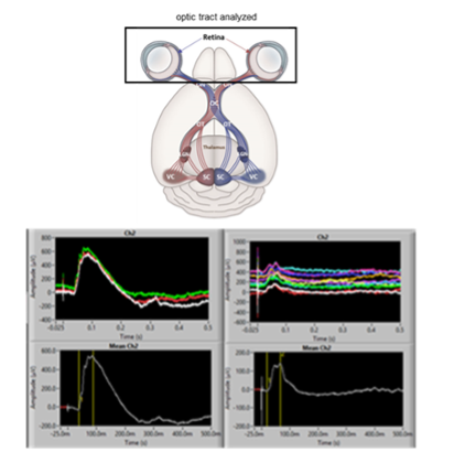
The major technologies that are available in the facility include: retinal electrophysiological tests in vivo and ex vivo, optical coherence tomography, pupillary light reflex, optomotry and fundus photographic. These tools vary in their invasiveness, complexity and optic tract analyzed.
