CORE FACILITY
Advanced Histopathology
Head: Nicolina Cristina Sorrentino
Experienced Technicians: Martina Sofia
Facility Group Mail: [email protected]


The analysis of morphological and pathological markers represents one of the fundamental readouts by which all preclinical and clinical studies are assessed. For this reason, the histopathological analyses of tissue samples should be performed with standardized, reproducible, and optimized experimental criteria.
The Advanced Histopathology Facility provides high quality histopathological services to the internal scientific community and to external research centers. The AHF also offers its experienced personnel for customized pathology services in order to meet the needs of scientists involved in basic, preclinical, and clinical research.
AHF Services provided:
- Tissue dissection and processing
- Automated paraffin embedding and OCT® embedding
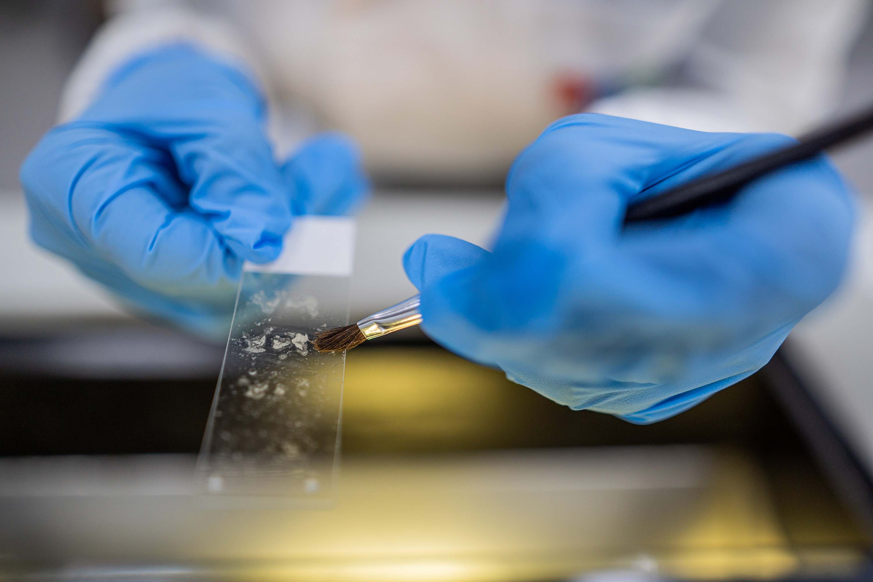
- Microtomy and cryostat sectioning of a wide variety of tissue samples
- Slide labeling
- Wide ranging histochemical staining (e.g. H&E, Masson trichrome, Toluidine blue, PAS, Cresyl violet, Alcian Blue, etc.)
- Innovative customized histological procedures to develop suitable approaches for:
- Automated detection of proteins, DNA and RNA in cells, animal and human tissues for single or multiplex immunofluorescence (IF) and immunohistochemistry (IHC) experiments
- Immunofluorescence (IF) experiments with 5 different fluorophores
- Immunohistochemical (IHC) experiments with 9 different chromogens
- Use of multiple primary antibodies raised in the same species, without cross talk
- Automated DNA/RNA detection through in situ hybridization (ISH) protocols
- Combined detection of DNA/protein & RNA/protein in the same tissue
- RNAscope technology for highly sensitive detection of RNA and miRNA
- Tunel assay in tissue samples
- co-localization analysis of proteins with fluorescent and chromogenic (translucent chromogens) staining.
The AHF also offers:
- Setting up and optimization of histological (IHC/IF) approaches for each scientific project by studying tissue samples characteristics, protein localization, antibody species and functionality.
- Validation of the best sequence of markers in multiplex experiments.
- Standardization of the IHC experiments and creation of a reference library of each antibody.
- Traceability of each histopathological, ISH, IF experiments performed.
- Practical training to free access to the equipment’s in the core facility like microtomes or cryostats.
- A large panel of titrated primary antibodies for human and mouse species available to the researchers.
- Delivery of protocols and microscope images of all histological experiments performed to the researchers
- Image Processing & analysis: AHF also offers support in processing and analysis of microscopic tissue images stained. The main tasks associated are the identification of different cell types and counting, the measurement of cellular parameters such as fluorescent intensity and area, morphometric analysis with specific software.
AHF Equipment:
EXCELSIOR AS AHSI (Thermo Fisher Scientific), this is an automated system for the dehydration and paraffin infiltration of human, plant, and animal tissues. Protocols are customisable according to each sample's needs.
EMBEDDING STATION (HistoCore Arcadia, LEICA), this station is used to position and embed tissues in molten paraffin wax to form paraffin blocks for use in microtome sectioning.
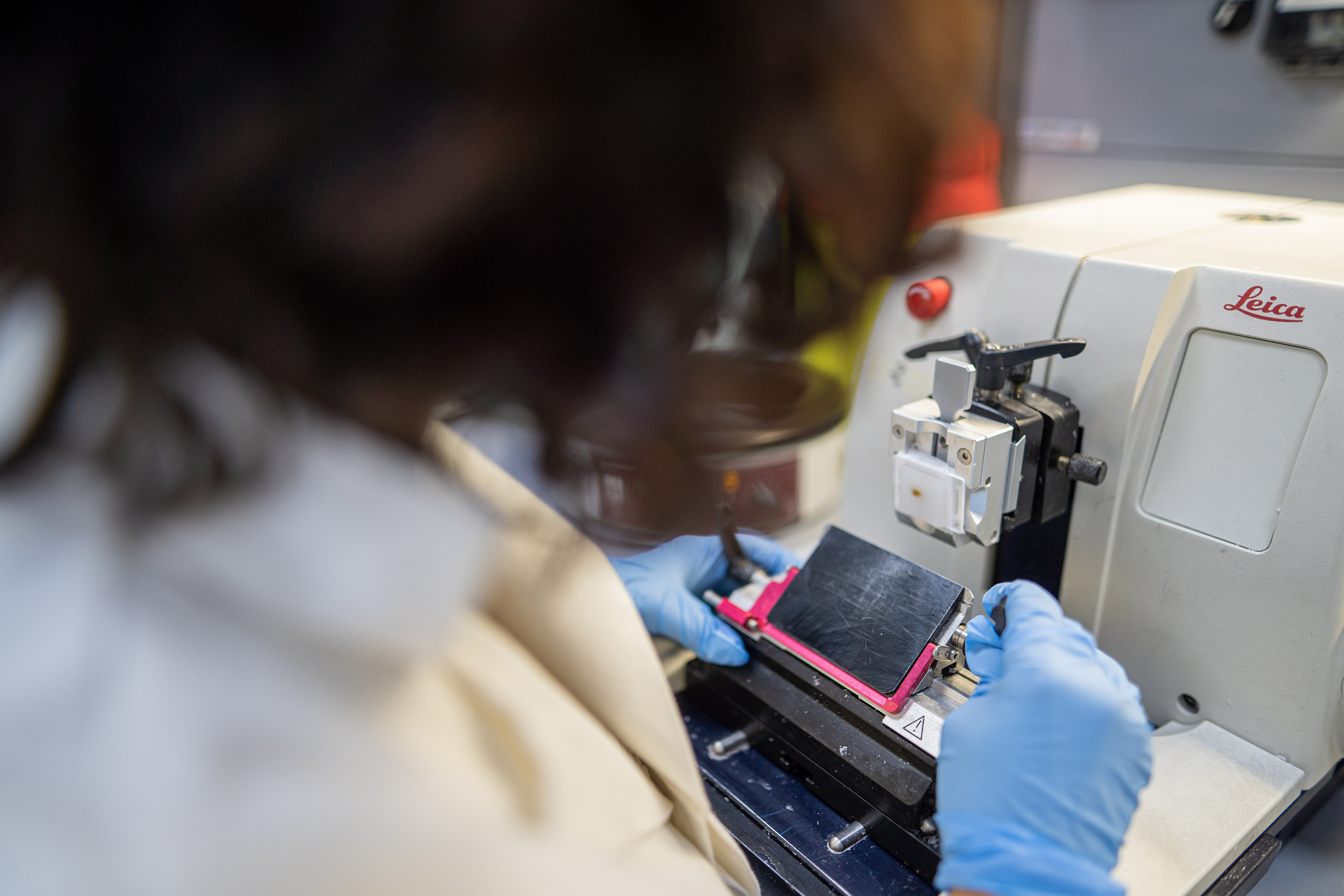
MICROTOMES (LEICA). We have both manual and semi-motorized rotary microtomes. Microtomes slice paraffin embedded tissues into sections as thin as 1μm. These sections are then mounted onto microscope slides, for use in histochemical, immunohistochemical, and immunofluorescence staining applications.
CRYOSTATS (LEICA). The cryostat is used for rapid freezing and sectioning of tissue samples. Fresh tissue samples are frozen with OCT embedding medium to provide rigidity and support for sectioning. Fresh muscle and bone samples can be sectioned by cryostats.
VENTANA DISCOVERY ULTRA (ROCHE). The Discovery Ultra enables high quality, highly reproducible chromogenic and fluorescence immunostaining. It is a fully automated system with enhanced protocol flexibility to process single or multiplex slides for IHC, IF, and ISH experiments in cells and tissues.
