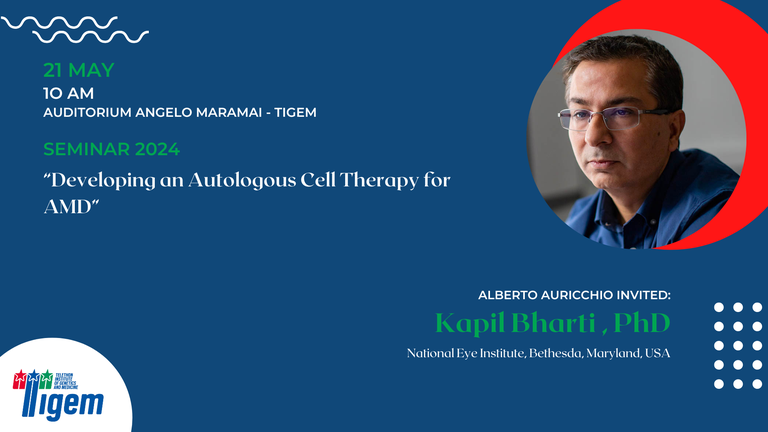
Kapil Bharti , PhD - "Developing an Autologous Cell Therapy for AMD"
- When May 21, 2024 from 10:00 AM to 11:15 AM (Europe/Berlin / UTC200)
- Where Tigem, Auditorium Angelo Maramai
- Contact Name Alberto Auricchio
- Contact Phone 08119230659
-
Add event to calendar
iCal

- https://www.tigem.it/newsroom/seminars/kapil-bharti-phd-developing-an-autologous-cell-therapy-for-amd-1
- Kapil Bharti , PhD - "Developing an Autologous Cell Therapy for AMD"
- 2024-05-21T10:00:00+02:00
- 2024-05-21T11:15:00+02:00
Kapil Bharti , PhD
National Eye Institute
Bethesda, Maryland
USA
Short CV
Abstract
The advanced stage of age-related macular degeneration (AMD) - geographic atrophy (GA) leads to irreversible vision loss and often manifests in people above 60 years of age. AMD is thought to initiate by the dysfunctional retinal pigment epithelium (RPE) monolayer - resulting in sub-RPE protein-rich drusen deposits. RPE sits between photoreceptors and choroidal capillaries, providing nutrients and support necessary for maintaining the homeostatic unit at the back of the eye. The GA stage is characterized by RPE atrophy, photoreceptor cell death, and the loss of chorio-capillary bed. Currently, no treatment is available to improve vision for the late stage of GA patients. Here, we developed an autologous cell replacement therapy for treating GA patients. We used induced pluripotent stem cells (iPSC) reprogrammed from CD34+ cells isolated from PBMC collected from patients' blood. iPSCs are differentiated into pure RPE cells using a protocol developed in our lab. The RPE cells are matured on a biodegradable polylactic co-glycolic acid (PLGA) scaffold for five weeks as a tissue patch. Quality control assays confirmed the iPSC-RPE patch's purity, maturity, and functionality. Pre-clinical studies were conducted in rats and pigs to demonstrate the safety and efficacy of the iPSC-RPE patch. Immune-compromised rats transplanted with a 0.5 mm iPSC-RPE patch showed no signs of tumor formation after nine months, confirming the safety profile. To test local safety and efficacy in a large animal model, we laser-ablated the RPE monolayer in the visual streak of pig eyes and, after 48 hours, transplanted the iPSC-RPE patch. Optical coherence tomography (OCT) that measures retinal anatomy confirmed the integration of the patch in the subretinal region. Multi-focal electroretinogram (ERG) that measures retina function showed that the electric response of the retinal layers over the area of patch was much higher than the lasered area without the implant. This work was cleared by the FDA for a Phase I/IIa clinical trial to test the safety and feasibility of an autologous iPSC-RPE patch in AMD patients with GA. The Phase I/IIa trial is currently ongoing at the National Eye Institute at NIH.
